Artroskopia stawu skokowego
BCM
5 grudnia 2023
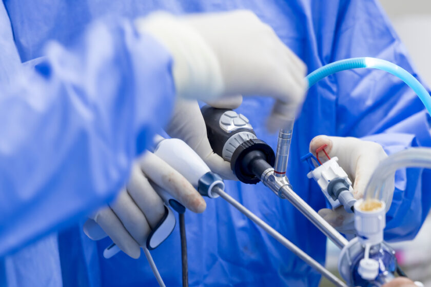
A rtroskopia to technika operowania stawów przez „dziurkę od klucza”. Instrumentarium zawierające miniaturową kamerę wideo (artroskop) jest wprowadzane do stawu, razem z innymi, miniaturowymi narzędziami celem naprawy uszkodzenia. Pozwala to chirurgowi na precyzyjną ocenę problemu oraz natychmiastową naprawę podczas jednego zabiegu operacyjnego.
Zabiegi artroskopowe w obrębie stawu skokowego z reguły wymagają jednej doby pobytu w szpitalu. Znieczulenie może być ogólne lub przewodowe (dokręgosłupowe)-w zależności od decyzji anestezjologa. Celem wprowadzenia instrumentów operacyjnych do stawu są wykonywane niewielkie nacięcia skórne w okolicy stawu. Po wprowadzeniu instrumentów staw wypełniany jest roztworem sterylnej soli fizjologicznej, pod kontrolą precyzyjnej pompy. Po wykonaniu naprawy (rekonstrukcji) uszkodzeń wewnątrzstawowych następuje założenie szwów oraz założenie opatrunku. W przypadkach rekonstrukcji niestabilności stawu wymagane jest zastosowanie stabilizatora stawu skokowego. Pacjent następnego dnia jest wypisywany do domu z precyzyjnymi informacjami odnośnie dalszych kontroli, rehabilitacji oraz okresu chodzenia w asekuracji kul łokciowych.
Przyczyny
Najczęstsze przyczyny wykonywania zabiegów artroskopii stawu skokowego to konflikty na podłożu tkanek miękkich przedniego lub tylnego przedziału stawu skokowego, uszkodzenia chrzęstno-kostne kości skokowej, ciała wolne, złamania w obrębie stawu skokowego, artrodeza (usztywnienie) stawu, niestabilności stawu skokowego, infekcyjne zapalenie stawu skokowego oraz zwłóknienie stawu czyli artrofibroza.
Objawy
Typowe objawy uszkodzeń stawu skokowego to dolegliwości bólowe (szczególnie podczas aktywności sportowej), obrzęki, ograniczenie ruchomości stawu, niestabilność, uczucie przeskakiwania w stawie, miejscowy wzrost temperatury.
Diagnoza
W trakcie konsultacji przedoperacyjnej po zebraniu wywiadu (historii schorzenia, dotychczasowego leczenia) lekarz wykonuje badanie kliniczne. To zestaw specjalistycznych testów określających prawdopodobną przyczynę dolegliwości pacjenta. Kolejny etap to wykonanie badań obrazowych-rtg, tomografii komputerowej, a obecnie najczęściej wykonywanym badaniem jest wysokopolowy rezonans magnetyczny. Dopiero wszystkie elementy diagnostyczne pozwalają na precyzyjne rozpoznanie.
Leczenie
Technika operacji artroskopowych przedniego i tylnego przedziału stawu skokowego umożliwia resekcję (wycięcie) masywnych zwłóknień, przerośniętej błony maziowej, usunięcie interponujących wyrośli chrzęstno-kostnych kości piszczelowej lub skokowej, usunięcie ciał wolnych, naprawę uszkodzeń powierzchni stawowych, w szczególności wydzielającej martwicy chrzęstno-kostnej (OCD) kości skokowej. Wykonujemy artroskopowo usztywnienie czyli artrodezę stawu w przypadku nasilonych zmian zwyrodnieniowych. Pod kontrolą artroskopu wykonuje się zespolenia złamań śródstawowych okolicy stawu skokowego, gdzie istnieje konieczność precyzyjnego odtworzenia powierzchni chrzęstnych. W przypadkach niestabilności stawu, szczególnie u pacjentów aktywnych sportowo wykonujemy rekonstrukcje więzadłowe. Artroskopię stawu skokowego wykorzystujemy także w przypadkach infekcyjnych.
Okres pooperacyjny
Pacjent spędza dobę po zabiegu operacyjnym w szpitalu. Następnego dnia, po zmianie opatrunku, usunięciu drenów ze stawu, kontroli czucia i ukrwienia kończyny operowanej następuje pionizacja pacjenta oraz nauka chodzenia o kulach pod nadzorem fizjoterapeuty. Po otrzymaniu niezbędnych wskazówek dotyczących dalszego postępowania oraz odbiorze dokumentacji medycznej pacjent opuszcza szpital. Dalsze leczenie-kontrole u lekarza prowadzącego oraz konsultacje rehabilitacyjne odbywają się w trybie ambulatoryjnym.
W Szpitalu Zakonu Bonifratrów artroskopie stawu skokowego wykonuje zespół specjalistów w dziedzinie ortopedii i traumatologii:
- lek. Stanisław Szymanik-Kierownik Oddziału Diagnostyczno-Leczniczego
- lek. Michał Latos
- lek. Michał Starmach