Artroskopia stawu kolanowego
BCM
12 lutego 2024
Zastosowanie artroskopii zrewolucjonizowało ortopedię. Dlaczego? Artroskopia stawu kolanowego polega na wprowadzeniu małej kamery do stawu, co pozwala lekarzowi zobaczyć wnętrze stawu, wykonując jedynie małe nacięcie.
Kolano było pierwszym stawem, w którym artroskop był powszechnie stosowany zarówno do diagnozowania problemów, jak i naprawy uszkodzeń. Artroskopia kolana pozwala bowiem na połączenie diagnostyki i leczenia – oczywiście z pożytkiem dla pacjenta.
Z czego zbudowany jest staw kolanowy?
Staw kolanowy to staw pomiędzy kością udową (powyżej), a kością piszczelową (poniżej). Z przodu stawu znajduje się rzepka. Duże mięśnie uda pozwalają nam poruszać kolanem. Staw kolanowy jest otoczony torebką stawową. Kapsuła ta jest utworzona przez więzadła kolana, tkankę łączną i tkankę maziową. Podczas artroskopii torebka stawowa wypełniona zostaje sterylną solą fizjologiczną, przez co w pewnym sensie artroskopia stawu kolanowego jest operacją „podwodną” gdyż wszelkie interwencje prowadzone są w środowisku soli fizjologicznej. Artroskopia stawu kolanowego pozwala lekarzowi zobaczyć prawie wszystkie struktury stawu, w tym: powierzchnie piszczeli, kości udowej i rzepki, obie łąkotki, więzadło krzyżowe przednie i tylne, wyściółkę maziową. Pomiędzy powierzchniami stawowymi, po obu stronach znajdują się łąkotki- przyśrodkowa i boczna.
Łąkotki pełnią funkcję ochronną, amortyzującą i stabilizującą kolano. Powierzchnie stawowe pokrywa chrząstka stawowa – gładki, śliski materiał. Chrząstka stawowa umożliwia wzajemne przesuwanie się powierzchni stawu bez uszkodzenia żadnej z nich. Więzadła to wytrzymałe pasma tkanki łączące końce kości razem. Więzadło krzyżowe przednie (ACL) znajduje się w środku stawu kolanowego, gdzie biegnie od tylnej części kości udowej, aby połączyć się z przednią częścią kości piszczelowej.
ACL biegnie przez specjalne wcięcie w kości udowej zwane wcięciem międzykłykciowym i przyczepia się do kości piszczelowej. ACL ma za zadanie zablokowanie nadmiernych ruchów piszczeli do przodu względem kości udowej oraz stabilizację kolana podczas ruchu. Więzadło krzyżowe tylne (PCL) znajduje się blisko tylnej części stawu kolanowego. Przyczepia się do tylnej części kości udowej i tylnej części piszczeli, do tyłu od ACL. PCL jest podstawowym stabilizatorem kolana i głównym kontrolerem tego, jak daleko w tył porusza się piszczel względem kości udowej.
W jakim celu wykonywana jest artroskopia stawu kolanowego?
Obecnie artroskopia kolana jest stosowana nie tylko do potwierdzenia diagnozy, ale przede wszystkim do wykonywania szerokiej gamy różnych zabiegów w stawie kolanowym, takich jak usunięcie ciał wolnych, usunięcie lub naprawa uszkodzonej łąkotki, rekonstrukcja zerwanych więzadeł, naprawa chrząstki stawowej, zaopatrzenie uszkodzeń rzepki. Obraz artroskopu jest powiększony i pozwala lekarzowi dokładniej przyjrzeć się strukturom stawu.
Minimalizacja nacięć i precyzja ruchów powoduje mniejsze uszkodzenie zdrowej tkanki i może skrócić proces gojenia. Pamiętać należy jednak, że artroskop jest tylko narzędziem. Artroskopia stawu kolanowego zasadniczo kończy się powrotem do sprawności, jednak wynik końcowy zależy od tego, co jest zasadniczym problemem i co można z nim zrobić. Równie istotną rolę co sam zabieg odgrywa również rehabilitacja po operacji.
Artroskopia kolana – co warto wiedzieć przed operacją?
Ty i Twój lekarz powinniście wspólnie podjąć decyzję o operacji. Powinieneś dowiedzieć się jak najwięcej o procedurze. Jeśli masz obawy lub pytania – koniecznie przedyskutuj swoje wątpliwości z lekarzem. Po podjęciu decyzji o operacji należy wykonać kilka kroków. Twój lekarz może zlecić dodatkowe badania i konsultacje. Wszystko po to, by upewnić się, że jesteś w stanie poddać się operacji i zminimalizować ryzyko powikłań. W dniu zabiegu zostaniesz przyjęty na Oddział Diagnostyczno – Leczniczy wcześnie rano. Nie powinno się jeść ani pić po północy poprzedniej nocy.
Na czym polega artroskopia stawu kolanowego?
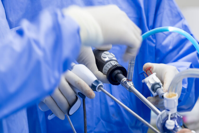
Przed zabiegiem pacjent zostanie poddany znieczuleniu ogólnemu lub znieczuleniu podpajęczynówkowemu. Decyzję o rodzaju znieczulenia podejmuje lekarz anestezjolog na podstawie rozmowy przed operacją – tzw. kwalifikacji do znieczulenia, podczas której wybierana jest najbardziej optymalna metoda znieczulenia. Artroskopia stawu kolanowego rozpoczyna ortopeda wykonując dwa małe nacięcia w kolanie zwane portalami. Portale te są miejscem, przez które artroskop i instrumenty chirurgiczne wprowadzane są do kolana. Artroskop jest podłużnym narzędziem o średnicy około 5 milimetrów.
Światłowód wewnątrz artroskopu pozwala na podłączenie jasnego światła i kamery. Kamera wyświetla na ekranie obraz z wnętrza stawu kolanowego. Po zakończeniu zabiegu artroskopowe portale zostają zamknięte za pomocą szwów. Najczęściej w jednym z portali zostaje umieszczony tzw. dren czyli giętka rurka połączona z plastikową butelką aby w okresie pooperacyjnym zbierać nadmiar krwi, która pojawi się w kolanie. Dreny zazwyczaj usuwane są następnego dnia po operacji.
Artroskopia stawu kolanowego – co po operacji?
Artroskopia kolana jest zwykle wykonywana już w dniu przyjęcia, a następnego dnia, tuż po zmianie opatrunku pacjent wraca do domu. Nawet bardziej skomplikowane rekonstrukcje więzadeł wymagają krótkiego pobytu w szpitalu, aby lepiej nadzorować ból i monitorować sytuację. Kule są powszechnie stosowane po artroskopii kolana, aby w okresie rekonwalescencji wspomóc pacjenta w przemieszczaniu się oraz odciążyć operowaną nogę.
Pacjenci, którzy przeszli bardziej skomplikowaną operację rekonstrukcyjną, mogą być poinstruowani, aby nie obciążać operowanej kończyny przez kilka tygodni (co oznacza, że nie mogą przenosić ciężaru ciała na operowaną kończynę – chodzą z pomocą kul łokciowych). Po rekonstrukcjach więzadłowych często stosuję się dodatkową ortezę, dzięki której można kontrolować zakres ruchów w kolanie.
Artroskopia stawu kolanowego w Szpitalu Zakonu Bonifratrów wykonywana jest przez zespół specjalistów w dziedzinie ortopedii i traumatologii:
- lek. Stanisław Szymanik – Kierownik Oddziału Diagnostyczno-Leczniczego
- lek. Michał Latos
- lek. Michał Starmach