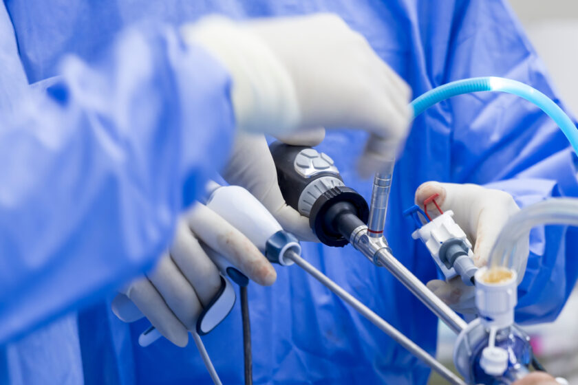
ARTHROSCOPIC RECONSTRUCTION OF THE POSTERIOR CRUCIATE LIGAMENT (PCL)
Introduction
The posterior cruciate ligament (PCL) is located to the back from the anterior cruciate ligament – just like the ACL, it connects the femur to the tibia. The structure prevents the so-called posterior tibial translation or moving the tibia backwards in relation to the femur, it also stabilizes the joint.
Causes
An injury of the posterior cruciate ligament is much rarer than that of the anterior cruciate ligament. The structure is much more durable than the anterior cruciate ligament. It is usually damaged when the bent knee is hit from the front, i.e. during traffic accidents or practicing competitive sports.
Symptoms
Like an injury of the anterior cruciate ligament, damage to the posterior cruciate ligament causes joint instability. In the acute phase, there is also swelling and pain, often with the limited mobility. The symptoms are significantly influenced by the condition of the remaining articular structures – menisci, cartilage and other ligaments, which are often also damaged during an injury.
Diagnosis
The initial diagnosis is often made after taking medical history and performing a clinical examination of the joint, it is confirmed by a diagnostic examination – magnetic resonance imaging of the knee.
Treatment
In most cases, the treatment is surgical. The chief aim of the operation is to restore the function of the PCL, i.e. to prevent excessive shifting of the tibia back against the femur – improving the stability of the knee.
Type of operation
The operation is performed by arthroscopy. In addition to three arthroscopic portals located in typical places, a fourth, longer incision (about 3-4 cm) is made below the knee – this is necessary to collect the tendons for transplantation.
When preparing the graft for reconstruction, the doctor assesses the joint, treats other injuries (for example, meniscus damage) and prepares the site for a new ligament – removes the remains of damaged PCL and carefully plans the location of the graft.
Then, using arthroscopic portals and the site after the tendon collection, the doctor drills canals in the femur and tibia and places the previously prepared graft in the appropriate position. After fixing the graft and controlling its function, the wounds are sutured, drains are placed and a sterile dressing is applied. A joint orthosis is usually put on already in the operating theater.
Postoperative period
In most cases, the day after surgery, the drains are removed and the patient gets out of bed with the help of elbow crutches. The operated limb is held in an orthosis. If everything is fine, the patient is discharged from the hospital. Rehabilitation should begin as soon as possible, because it is relevant after the reconstructive surgery. The therpay should start within 5 days of the procedure.
In the Szpital Zakonu Bonifratrów, reconstructions of the cruciate ligaments are performed by a team of specialists in orthopedics and traumatology:
Stanisław Szymanik, MD – Head of the Diagnostic and Treatment Department
Michał Latos, MD
Michał Starmach, MD
REHABILITATION