Biopsja mammotomiczna – małoinwazyjne leczenie łagodnych guzków piersi
BCM
5 grudnia 2023
G uzek w piersi, czy inne zmiany ogniskowe piersi są często wykrywane w trakcie samobadania przez pacjentki, rutynowych badań lekarskich lub badań obrazowych. Można je podzielić na zmiany nienowotworowe jak np. torbiele, nowotwory łagodne (gruczolakowłókniaki, brodawczaki, tłuszczaki) oraz nowotwory złośliwe (rak piersi). Zmiany łagodne (guzek piersi), nowotworowe i nienowotworowe występują częściej niż rak piersi i generalnie w młodszym wieku, jednak często postawienie właściwej diagnozy i wdrożenie odpowiedniego leczenia jest trudne.
Diagnoza
Diagnostyka zmian ogniskowych piersi opiera się na badaniach obrazowych i metodach biopsyjnych. Do najczęściej wykorzystywanych badań można zaliczyć USG, mammografię i rezonans magnetyczny, dzięki którym zobaczyć można guzek w piersi oraz inne zmiany. Metody te, uzupełniając się, pozwalają z dużym prawdopodobieństwem postawić właściwe rozpoznanie. Ostateczną diagnozę stawia się na podstawie badania histopatologicznego materiału pobranego z guzków w trakcie biopsji. Najczęściej wykorzystywaną metodą jest biopsja gruboigłowa, czyli biopsja mammotomiczna pozwalająca na pobranie wycinków z guza. Rzadziej w diagnostyce guzów piersi wykorzystuje się biopsję cienkoigłową lub chirurgiczną (wycinającą).
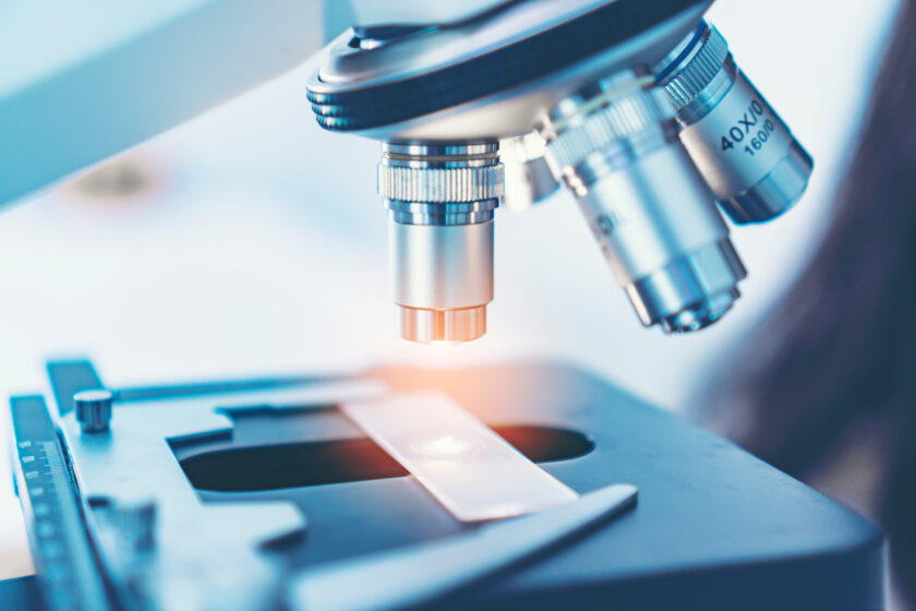
Co to jest biopsja mammotomiczna?
Biopsja mammotomiczna, a właściwie biopsja gruboigłowa wspomagana próżnią, pozwala zdiagnozować podejrzany guzek piersi. Biopsja mammotomiczna umozliwia w sposób minimalnie inwazyjny pobrać wycinki ze zmian podejrzanych oraz usunąć w całości łagodny guzek piersi. Procedura wykonywana jest najczęściej pod kontrolą USG, ale w wyspecjalizowanych centrach, może też być przeprowadzana pod kontrolą mammografii stereotaktycznej.
Biopsja mammotomiczna wykonywana jest prze pomocy zestawu złożonego z jednorazowej igły biopsyjnej z systemem przewodów, rękojeści pozwalającej sterować pracą igły oraz jednostki centralnej wytwarzającej podciśnienie i monitorującej pracę urządzenia. Zabieg jest wykonywany w warunkach ambulatoryjnych, w znieczuleniu miejscowym, w pozycji leżącej. Przez niewielkie nacięcie (około 2-3 mm) igła biopsyjna wprowadzana jest w okolicę miejsca, w którym jest guzek w piersi. Następnie fragment tkanki zasysany jest do wnętrza igły i odcinany przez poruszający się nóż. Wycinek dzięki systemowi przewodów transportowany jest na zewnątrz i po utrwaleniu przesyłany do oceny histopatologicznej. Po wycięciu guzka lub pobraniu wycinków igła jest usuwana i zakładany jest opatrunek uciskowy. Ze względu na niewielkie rozmiary rany skórnej nie ma konieczności zakładania szwów. Pacjentka po biopsji pozostaje zazwyczaj w obserwacji przez około 30 minut, a następnie może iść do domu. Aby zminimalizować ryzyko wystąpienia krwawienia i krwiaka opatrunek uciskowy powinien być utrzymany przez 24 godziny.
Biopsja mammotomiczna – powikłania
Biopsja mammotomiczna charakteryzuje się tym, że powikłania po niej zdarzają się rzadko i obejmują: krwiaki oraz łagodne bóle w miejscu wkłucia, omdlenia, zaburzenia gojenia się rany, powstanie keloidu oraz reakcje alergiczne na podany anestetyk lokalny. Bardzo rzadko zdarzają się krwawienia wymagające interwencji chirurgicznej. Wyjątkowo rzadko może dojść do przebicia ściany klatki piersiowej i powstania odmy opłucnowej.
Biopsja mamotomiczna pozwala na wysłanie fragmentu guzka w piersi do badania histopatologicznego w celu ostatecznego ustalenia rodzaju zmiany. Potwierdzenia łagodnego charakteru guza kończy proces leczniczy. W przypadku stwierdzenia nowotworu złośliwego pacjentki kierowane są do leczenia w wyspecjalizowanych centrach.
W Szpitalu Zakonu Bonifratrów biopsje mammotomiczną – małoinwazyjne leczenie łagodnych guzków piersi wykonują dr n. med. Tomasz Gach.
Świadczenie udzielane jest wyłącznie odpłatnie.